FIGURE 3.

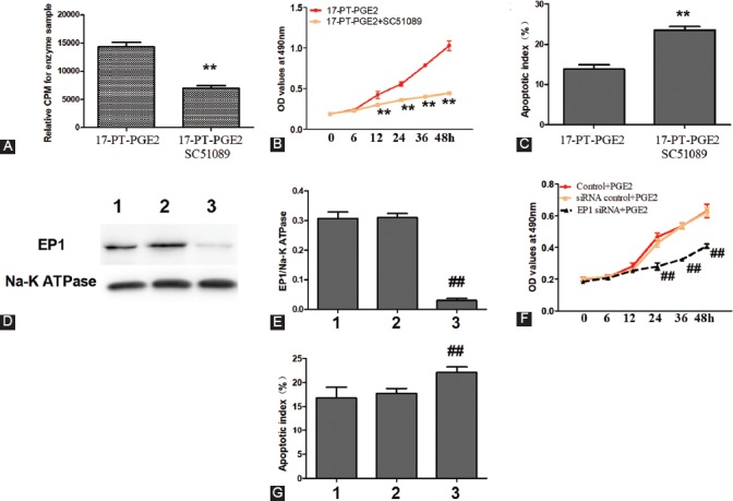
Blockade or silencing of the EP1 receptor decreased MG63 cell viability. MG63 cells were co-cultured with 17-PT-PGE2 (5 µM) and SC51089 (2 µM) for 48 hours. A) PKC activity assay. B) Cell viability was tested by MTT assay. C) MG63 cells were fixed and stained with DAPI and TUNEL. SC51089 significantly blocked 17-PT-PGE2-induced activation of the EP1/PKC pathway. In addition, MG63 cells exposed to 17-PT-PGE2 + SC51089 showed a decreased proliferation and increased apoptosis compared to MG63 cells exposed to 17-PT-PGE2 only. D-G) MG63 cells transfected with an EP1R-siRNA were exposed to PGE2 (5 µM) for 48 hours. The silencing of EP1 receptor suppressed PGE2-induced proliferation in MG63 cells. Knockout efficiency was measured by Western blotting; cell viability was tested by MTT assay; and apoptosis of MG63 cells was assessed by DAPI and TUNEL staining. 1: Control+PGE2; 2: siRNAControl+PGE2; 3: EP1R-siRNA+PGE2. Each sample was prepared in triplicate and all experiments were repeated three times. All bar/line graphs represent the mean ± SD. **p < 0.01 vs. 17-PT-PGE2; ##p < 0.01 vs. siRNA Control+PGE2. PGE2: Prostaglandin E2; PKC: Protein kinase C; EP1R-siRNA: siRNA targeting the EP1 receptor; siRNAControl: Non-silencing irrelevant RNA duplex.
