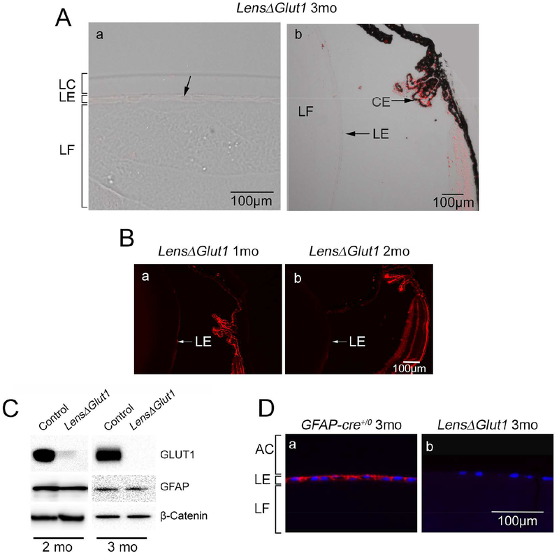
Figure 2: GLUT1 is deleted from LensΔGlut1 lens.
A. In the LensΔGlut1 lens, Slc2a1 expression was not detected in anterior (a) or equatorial (b) regions of the lens epithelium. In comparison, GLUT1 expression was retained in the ciliary epithelium. B. Immunofluorescence with GLUT1 in 1-month-old (a) and 2-month-old LensΔGlut1 lenses (b). The GLUT1 levels were low in the 1-month-old LensΔGlut1 mice and barely detectable in the 2-month-old mice. C. Western blot for GLUT1 in lysates of isolated lens epithelium from control and LensΔGlut1 mice at 2 months (left) and 3 months (right) of age. Note that the residual expression of GLUT1 in the 2-month-old LensΔGlut1 mouse is lost by 3 months of age. GFAP expression is unchanged in control versus LensΔGlut1 mice. ß-Catenin was used as a loading control. D. Immunohistochemistry for GLUT1 in lens epithelium of 3-month-old control (a) and LensΔGlut1 (b) mice. LC: lens capsule, LE: lens epithelium, LF: lens fiber cells, AC: Anterior chamber.

