Figure 1.
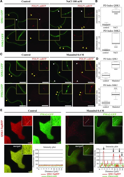
IMK2 and QSK1 are PM-associated LRR-RLKs that reorganize at plasmodesmata upon salt and mannitol treatments. A to D, Transient expression in N. benthamiana epidermal cells of IMK2-GFP and QSK1-GFP LRR-RLKs expressed under 35S promoter and visualized by confocal microscopy. In control conditions, the two LRR-RLKs localize exclusively at the PM and present no enrichment at plasmodesmata, which are marked by PDLP1-mRFP. Upon 100 mm NaCl (A and B) or 0.4 m mannitol (C and D) treatment (5–30 min), the two LRR-RLKs relocalize to plasmodesmata (arrowheads). Yellow-boxed regions are magnification of areas indicated by yellow arrowheads. Enrichment at plasmodesmata versus the PM was quantified by the PD index, which corresponds to the fluorescence intensity ratio of the LRR-RLKs at plasmodesmata versus the PM in control and abiotic stress conditions (see “Materials and Methods” for details and Supplemental Fig. S1). n = 4 experiments, 3 plants/experiment, 10 measures/plant. Two-tailed Wilcoxon statistical analysis, *P < 0.05; **P < 0.01; ***P < 0.001. E, Transient expression in N. benthamiana epidermal cells of QSK1-TagRFP and PMA2-GFP expressed under 35S promoter and visualized by confocal microscopy. Top surface view of a leaf epidermal cell showing the uniform and smooth distribution pattern of QSK1-TagRFP and PMA2-GFP at the PM under control conditions. Mannitol treatment causes a relocalization of QSK1-TagRFP, but not of PMA2-GFP, into microdomain-like structures at the PM on the upper epidermal cell surface. Intensity plot along the white dashed line visible on the confocal images. n = 2 experiments, 3 plants/experiment. Scale bars, 10µm.

