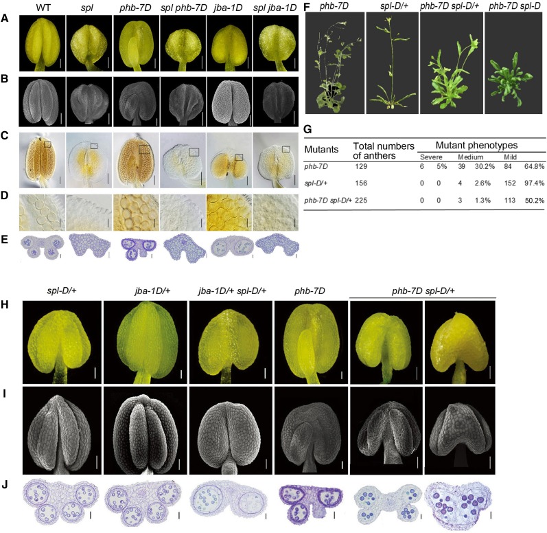
Figure 3.
Anther morphology and anatomy in the single and double mutants of SPL/NZZ, MIR166g, and PHB. A, Anthers of spl, jba-1D, and phb-7D mutants at stage 12. B, Scanning electron microscopy images showing the appearance of spl, jba-1D, and phb-7D anthers at stage 12. C, Whole-mount clearing images showing the internal structures of spl, jba-1D, and phb-7D locules at stage 12. D, Magnified images of the boxes in C showing the pollen grains in spl, jba-1D, and phb-7D locules at stage 12. E, Cross sections of spl, jba-1D, and phb-7D anthers. F, Plant phenotypes of phb-7D, spl-D/+, and jba-1D single and double mutants at flowering stage. G, The distribution of phb-7D spl-D/+ anthers with the mutant phenotypes of different severity. H, Anthers of phb-7D, spl-D/+, and jba-1D single and double mutants at stage 11 imaged with an anatomical microscope. I, Scanning electron microscopy images showing the appearance of anthers of the phb-7D, spl-D/+, and jba-1D single and double mutants at stage 11. J, Cross sections of the anthers of phb-7D, spl-D/+, and jba-1D single and double mutants at stage 11. Bars = 50 μm (A–C, H, and I) and 20 μm (D, E, and J).

