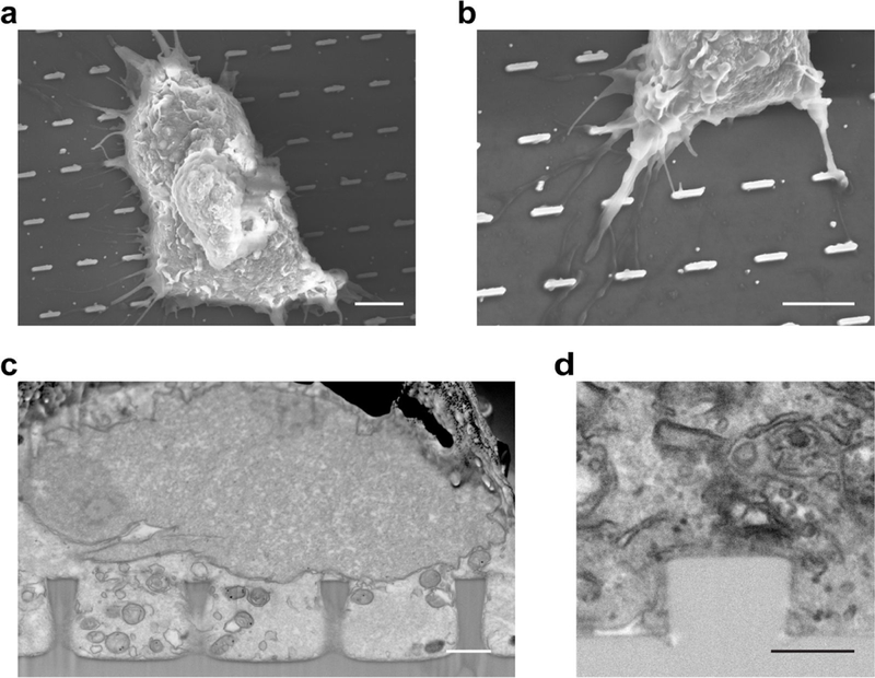
Fig. 11 |. FIB-SEM images of cells cultured on nanostructures.

a, SEM image of a cell growing on nanobars, b, SEM image of cell membrane extrusions on nanobars. The nanobars have 200 nm width, 2 μm length, and 1 μm height (a and b). c, Polished cross-sectional micrograph of a part of a cell on nanopillars. The nanopillars have 700 nm diameter and 1.6 μm height, d, Polished cross-sectional micrograph of a part of a cell on a nanopillar. The nanopillar has 700 nm diameter and 500 nm height. Scale bars, 5 μm (a and b), 1 μm (c); 500 nm (d).
