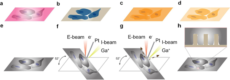
Fig. 9 |. Schematics of FIB-SEM workflow.

a, Cells cultured on a nanochip, b, Cells fixed in glutaraldehyde after culture, processed with the RO-T-O procedure and uranyl acetate to increase their contrast, c, Cells on a nanochip embedded in resin, d, resin excess removal and subsequent polymerization. To remove the resin excess, the sample is placed in a vertical position and gently wash them with ethanol, e, Cells on nanochip coated with Au. f, Pt deposition assisted by both E-beam and I-beam, g, I-beam induced cross sectioning, h, Imaging the cell-nanostructure interface after cross-sectioning.
