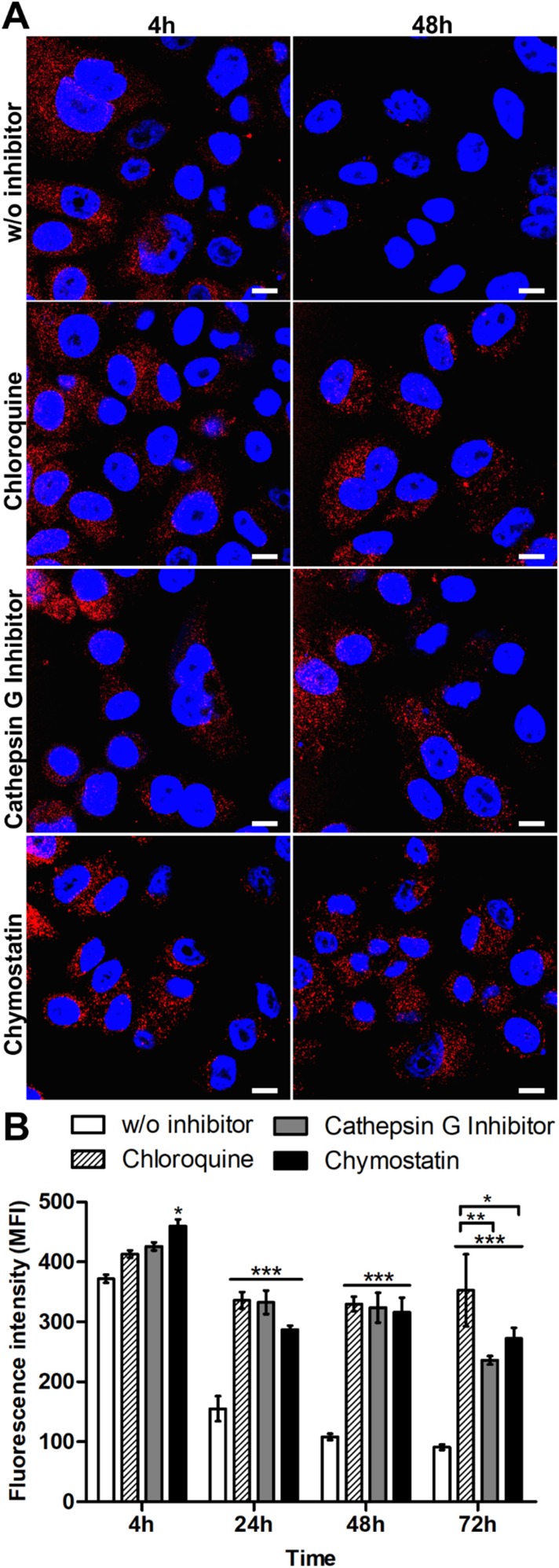
Figure 3.
Analysis of the intracellular processing of the silk spheres in SKBR3 cells. (A) The cells were incubated with ATTO647N-labeled H2.1MS1:H2.1MS2 spheres in the presence of lysosomal inhibitor chloroquine, cathepsin G inhibitor I or chymostatin for the indicated time periods at 37 °C. The silk particles were observed using CLSM. Red, spheres conjugated with ATTO647N; blue, nuclei stained with DAPI; scale bar: 10 μm. (B) The fluorescence intensity of the silk spheres was measured by the mean fluorescence value (±SEM) per cell (n=30). Statistical significance with p<0.001 (***), p<0.01 (**), p<0.05 (*).

