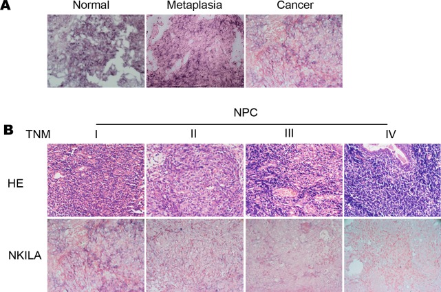
Fig 2. Low expression of NKILA is correlated with NPC progression.
Representative images (×400) (A) ISH for NKILA were performed using normal nasopharyngeal tissue, metaplasia nasopharyngeal tissue with atypical hyperplasia and NPC tissue. (B) H&E staining and ISH for NKILA were performed using paraffin-embedded NPC tissue with stage I to IV.

