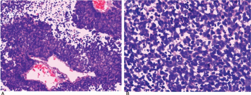
Figure 2.

At 100× magnification (A), we observed tumor cells to be densely distributed in patches or around blood vessels, with a papillary arrangement. At 400× magnification (B), the structure of the tumor cells was clearly visible. Less cytoplasm, as well as hyperchromatic and oval nuclei, were observed in tumor cells, accompanied by apoptosis and thanatosis. Diagnosis: small cell malignant tumor.
