Figure 1.
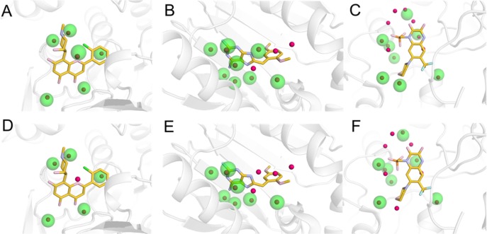
Examples of hydration sites derived from MD simulations and comparison to crystal waters for three protein–ligand complexes. Hydration sites derived in the presence (A-C) and absence (D-F) of the ligands are shown for muscle glycogen phosphorylase (A, D), heat shock protein 90-alpha (B, E), and glutamate receptor ionotropic, AMPA 2 (C, F). In (D-F), the cocrystallized ligands are shown, but they were not included in the MD simulations. Hydration sites and crystal waters are shown as transparent green and red spheres, respectively. The proteins are shown as gray cartoons, and the cocrystallized ligands are depicted in sticks.

