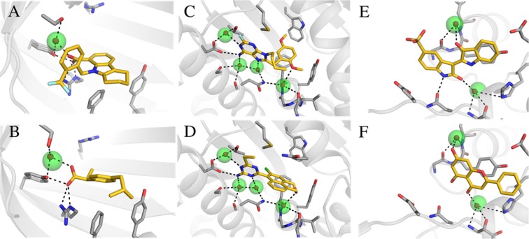
Figure 5.
Examples of hydration sites derived from the reference crystal structures (Table 1) that reproduced crystal waters forming hydrogen bonds with diverse ligands in other experimental structures. Two crystal structures for three different proteins are shown: (A, B) Fatty acid binding protein adipocyte (PDB codes: 3fr4 and 3p6h); (C, D) Heat shock protein 90-alpha (PDB codes: 1uyh and 3b28); (E, F) Muscle glycogen phosphorylase (PDB codes: 1uzu and 3ebo). Hydration sites and crystal waters are shown as transparent green and red spheres, respectively. The proteins are shown as gray cartoons with selected residues and the cocrystallized ligands in sticks. Hydrogen bonds are depicted as black dashed lines.

