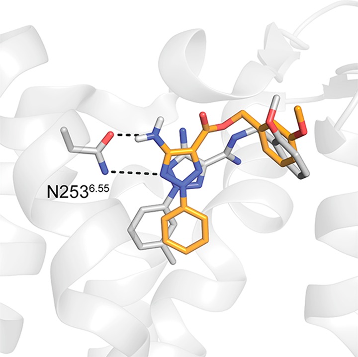
Figure 5.

Comparison of the predicted binding mode of compound 3 to a crystal structure of a related antagonist in complex with the A2AAR. The dual-target ligand 3 is shown as sticks with orange carbon atoms. The cocrystallized antagonist (PDB code 5UIG(46)) is depicted in sticks with gray carbon atoms. The A2AAR is depicted as gray cartoons. Hydrogen bonds are shown as black dashed lines.
