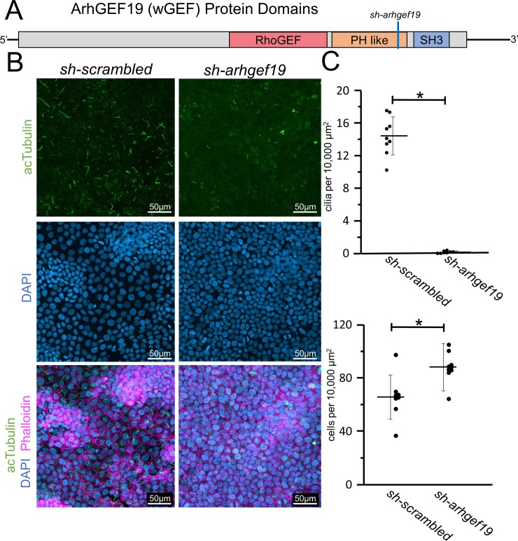
Fig 5. Arhgef19 knockdown results in loss of primary cilia in MDCKII cells.
A) Diagram of the domains within the Arhgef19 (WGEF) protein. The corresponding position of the shRNA is marked with a blue line. B) MDCKII cells were infected with either a control construct or a construct that targets Arhgef19 and then polarized on transwell filters. Cells were stained with acetylated α-Tubulin antibody (acTubulin) to visualize primary cilia (green), DAPI to label nuclei (blue), and phalloidin to label F-actin (magenta). Confocal imaging was used to analyze the effects Arhgef19 depletion upon primary ciliogenesis. Scale bars equal to 50 μm. C) The number of cilia was counted and cell numbers were quantified as described in S5 Fig. * indicates p < 0.05 as compared to sh-scrambled. Error bars are shown as ± SD and black dots indicate value of each image quatified.

