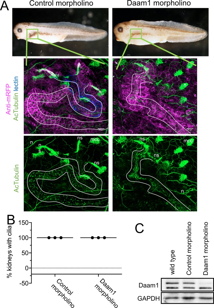
Fig 6. Daam1-depletion does not cause absence of cilia within Xenopus embryonic kidneys.
Because knockdown of Daam1 in Xenopus kidney leads to kidney defects, we analyzed the effect of Daam1 knockdown on renal ciliogenesis. We injected Daam1 or Standard (Control) morpholino in combination with a membrane-tagged red fluorescent protein (mRFP) mRNA as a lineage tracer, into a Xenopus blastomere fated to the nephric anlagen. Embryos were fixed at stage 39–40 and stained with an antibody against acetylated α-Tubulin (acTubulin) to label cilia (green), anti-mRFP lineage tracer (magenta) and lectin to label the proximal tubule (blue). A) Stereoscope brightfield imaging shows the gross morphology of Control and Daam1-morpholino injected embryos. Confocal fluorescent imaging of boxed regions (green) shows magnified views of corresponding kidneys. Kidney tubules displaying primary cilia are outlined in white. Neurons (n), multiciliated epidermal cells (mcc) and multiciliated cells within nephrostomes (ns) are immunostained with acetylated α-Tubulin (acTubulin) antibody. Scale bars equal to 20 μm. B) The graph represents the percentage of Control (n = 32 embryos) and Daam1-depleted (n = 34 embryos) kidneys with primary cilia. C) Western blot showing Daam1 protein expression levels in uninjected wild-type, control and Daam1 morphant embryos.

