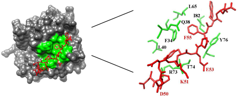
Figure 1:
An X-ray crystal structure of human PAR1 fragment (49–57) bound to thrombin (PDB code 3LU9). The thrombin surface is rendered in gray. The PAR1 residues are labeled as red sticks whereas ABE I residues situated < 4 Å from PAR1 are shown as green sticks or green surfaces. Residues later selected for 1H,15N-HSQC NMR titration studies are highlighted in red.

