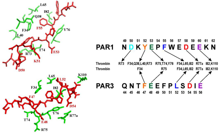
Figure 7:
Comparing interactions between thrombin ABE I (30 and 70 loop), PAR1 (49–62), and PAR3 (44–56). Crystal structures of thrombin in complex with human PAR1 (49–57) [PDB 3LU9] and murine PAR3 (44–56) [PDB 2PUX] were employed as structural guides. Since PAR1 (58–62) was unresolved by crystallography, proposed interactions with thrombin residues are provided for PAR1 D58 and E60. The PAR fragments target similar thrombin ABE I regions but to varying extents.

