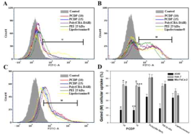
Figure 4.
Cellular uptake of polyplexes in (A) A549, (B) Huh-7, and (C) Mia PaCa-2 cells. The YOYO-1 stained pDNA were formed the polyplexes with PCDP, poly(CBA-DAH), PEI 25 kDa, and Lipofectamine® at weight ratio based on pDNA (1 µg), respectively. (D) The cellular uptake % of quantification of cell internalization measured by FACs. Results are represented as mean ± SD. (n=3). *<0.05, **P < 0.01, ***P <0.001 versus PEI 25 kDa.

