Fig. 3.
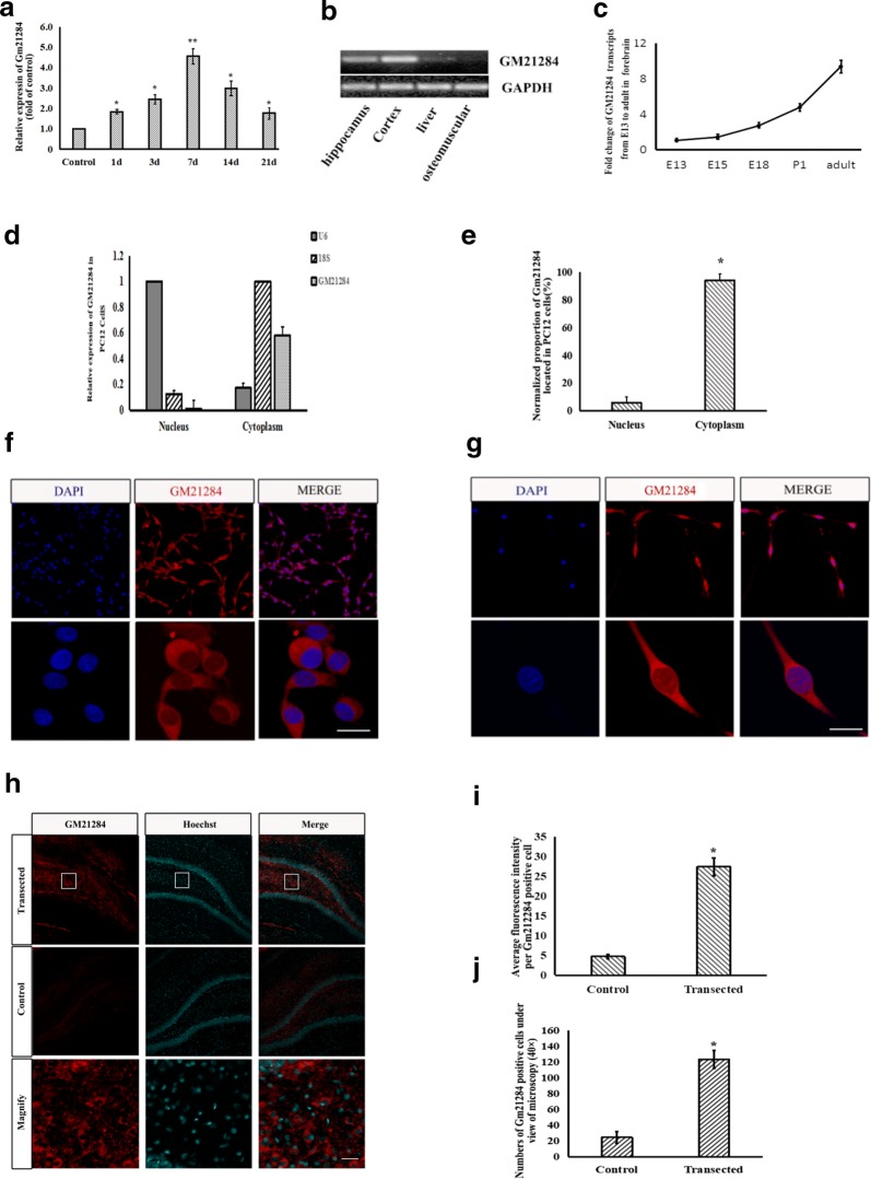
Expression and location of Gm21284. a RT-PCR detected Gm21284 expression in hippocampal niche after fimbria–fornix transection at different time points, *: vs. control group, P < 0.05; **: vs. control group, P < 0.01. b RT-PCR detected Gm21284 in tissues originated from different germinal layer. c RT-PCR detected Gm21284 in forebrain cortices of E13, E15, E18, and P1 and in adult rats. d, e RT-PCR showed that the expression of Gm21284 in cytoplasm of PC12 cells was higher than that in nucleus, *: vs. nucleus, P < 0.05. f FISH assay showed Gm21284 mainly localized in cytoplasm of PC12 cells. g FISH assay showed Gm21284 localized mainly in the cytoplasm of hippocampal neurons. h FISH labeled Gm21284 positive cells in the dentate gyrus of the hippocampus. i FISH showed that the average fluorescence intensity of Gm21284 positive cells in transected side was higher than the control side in DG after fimbria–fornix transection at 7 days, *: vs. control group, P < 0.05. j Confocal microscopy (×40) showed that the number of Gm21284 positive cells on the transected side was higher than the control side in DG after fimbria–fornix transection at 7 days, *: vs. control group, P < 0.05. Scale bar = 50 μm

