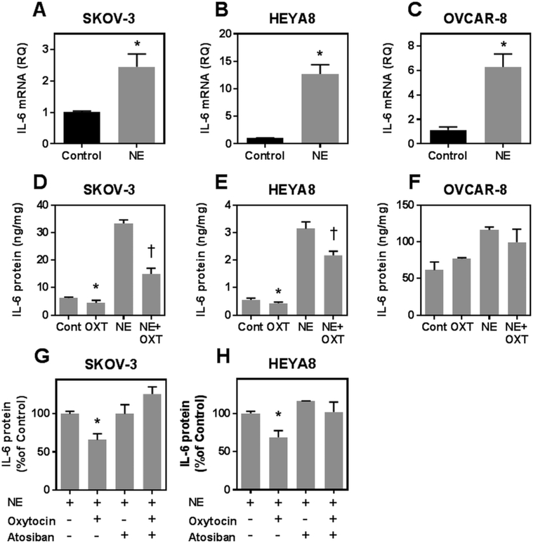
Figure 3.
Effect of oxytocin on ovarian tumor cell interleukin-6 secretion under basal and stimulated conditions. Panels A-C show interleukin-6 mRNA expression after exposing cells to control medium or medium with 10 μM norepinephrine (NE) for 6 hours. Panels D-F show interleukin-6 (IL-6) secretion from different cell lines following incubation without (Control) or with 1 nM oxytocin (OXT) under basal or NE stimulated conditions. All data are expressed as the mean ± SD of 3 or 4 dishes and representative of at least two similar experiments. * and † indicate p< 0.05 by Students t-test compared to control or NE stimulated conditions, respectively. Panels G and H show IL-6 secretion from SKOV-3 and HEYA8 cells treated with 10 μM NE for 6 hours alone or in the presence of 1 nM OXT, 10 nM Atosiban or OXT and Atosiban simultaneously as indicated. Data the mean ± SD of 3 dishes expressed as % IL-6 secretion relative to cells treated with NE (Control condition) and * indicates p< 0.05 by Students t-test compared to control.

