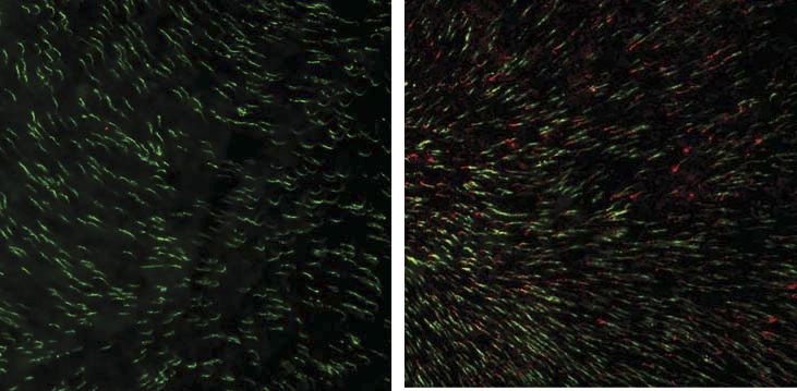
Figure 4.

AAV-delivered OPN1LW is expressed in Opn1mw−/− dorsal cones when treated at 15 months of age and tested at 2 months postinjection. A representative image of an Opn1mw−/− retinal flat mount immunostained with OPN1LW (red) and PNA (green) showing that OPN1LW is expressed in dorsal retinal cones and localized to the tip of PNA staining in the treated eye (right). In contrast, no OPN1LW expression was detected in the untreated contralateral eye (left).
