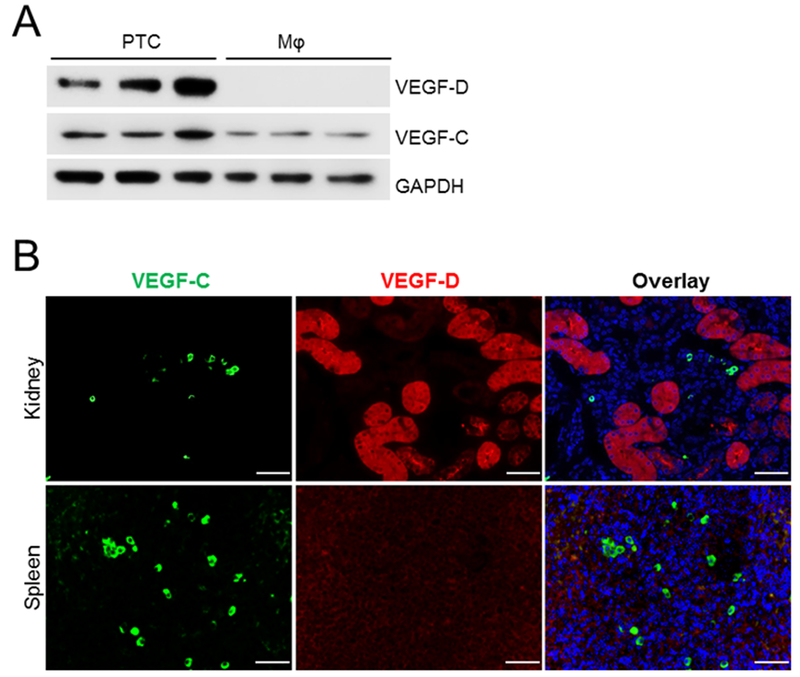
Figure 9. Tubular cells are major source of VEGF-D while both macrophages and tubules contribute to VEGF-C production.
(A) PTC and BMDM were harvested and their proteins were examined for VEGF-C and VEGF-D content via Western blotting. (B) Spleen and kidneys were stained for VEGF-C and VEGF-D. Immunofluorescence staining reveals macrophages that only express VEGF-C and renal tubules that express both VEGF-C and VEGF-D. Images are representative three independent experiments. Scale bar= 50 μm.

