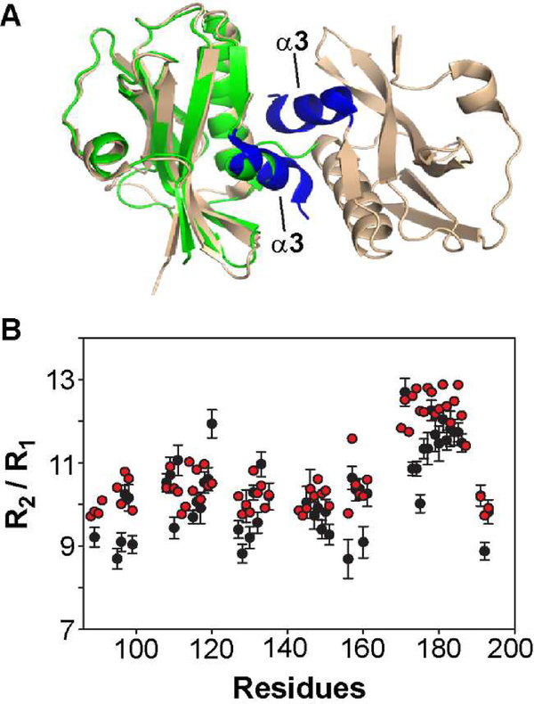
Figure 2.
1918 NS1-ED exists as a monomer in solution. (A) Crystal structure of 1918 NS1-ED W187A (PDB ID: 6DGK). The α3-α3 dimeric interface is shown in blue. For comparison, NMR structure of 1918 NS1-ED W187R (shown in green) is superimposed to chain A of the crystal structure. (B) The NMR R2 / R1 ratio as a function of primary structure of 1918 NS1-ED. The experimental and HYDRONMR-calculated values of conformationally rigid residues are shown in black and red circles, respectively.

