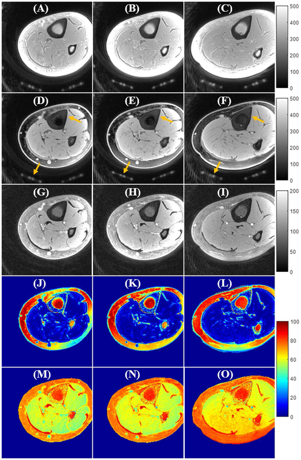
Figure 5.
In vivo tibia UTE-Cones imaging (from a 35-year-old volunteer) using excitations with a single hard pulse (A–C), the proposed soft-hard water excitation pulse (D–F), and the conventional FatSat module (G–I). Fat was well suppressed by both the proposed soft-hard pulse and the FatSat module. The cortical bone and coil elements (indicated by yellow arrows in D–F) were much better preserved in the soft-hard excitation images (D–F) compared with FatSat images (G–I). The SSR colormaps (soft-hard pulse: J–L; FatSat module: M–O) also suggest that there were almost no signal attenuations for either short or long T2 tissues when using the proposed soft-hard pulse for excitation. In comparison, there were strong signal attenuations for the water signals in the FatSat UTE-Cones images, especially for the short T2 tissues.

