
Figure 4.
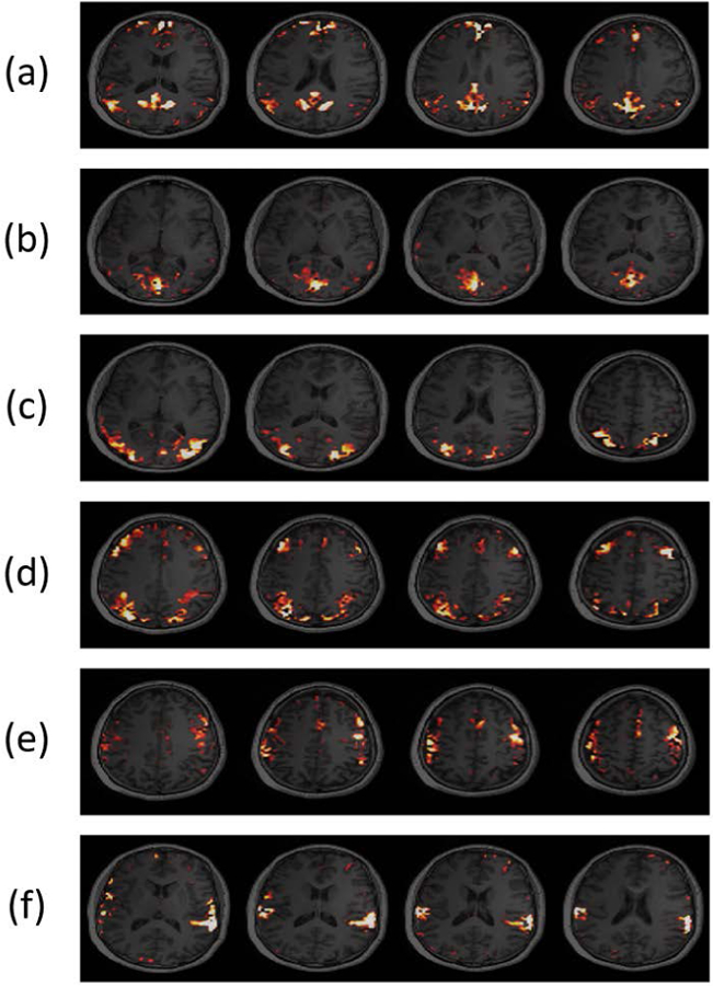
Activation maps obtained from a 6.5-min resting-state fMRI/MRSI scan using the proposed method. The maps showed spatial patterns for: (a) default mode network (DMN), (b) medial visual network (MVN), (c) lateral visual network (LVN), (d) executive control network (ECN) (e) sensorimotor network (SMN), and (f) auditory network (AN), respectively. These resting-state network structures are consistent with those in the literature (51–54).