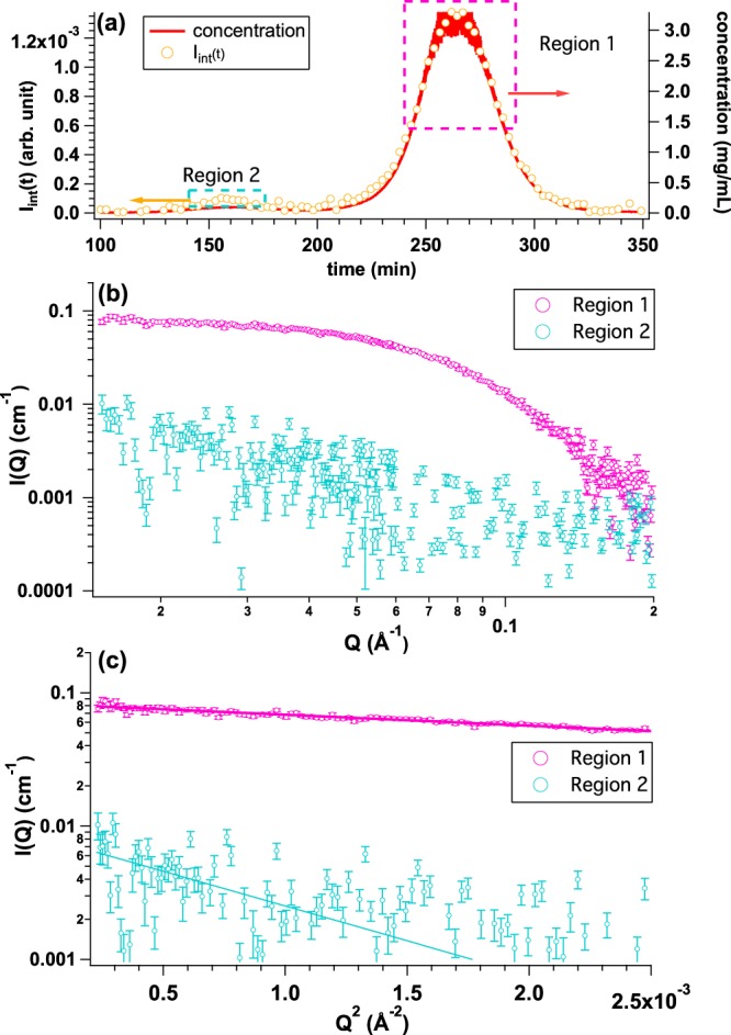
Figure 4.

(a) Time evolutions of integrated SAXS intensity (Iint(t)) (yellow circle) and concentration calculated from Uc(t) (UV-SAXS-cell: red line) by injecting OVA solution with the initial concentration and the volume of 5.0 mg/mL and 500 μl, respectively. We selected two regions; Region 1 (peak at t = 265 min: highlighted by pink dotted rectangle) and Region 2 (peak at t = 155 min: highlighted by light blue rectangle) for obtaining averaged SAXS profiles. (b) Averaged SAXS profiles over Region 1 (pink circle) and Region 2 (light blue circle), respectively. (c) Guinier plots of averaged SAXS profiles from Region 1 (pink, Rg = 23.9 ± 0.4 Å) and Region 2 (light blue, 63.8 ± 28.4 Å), respectively.
