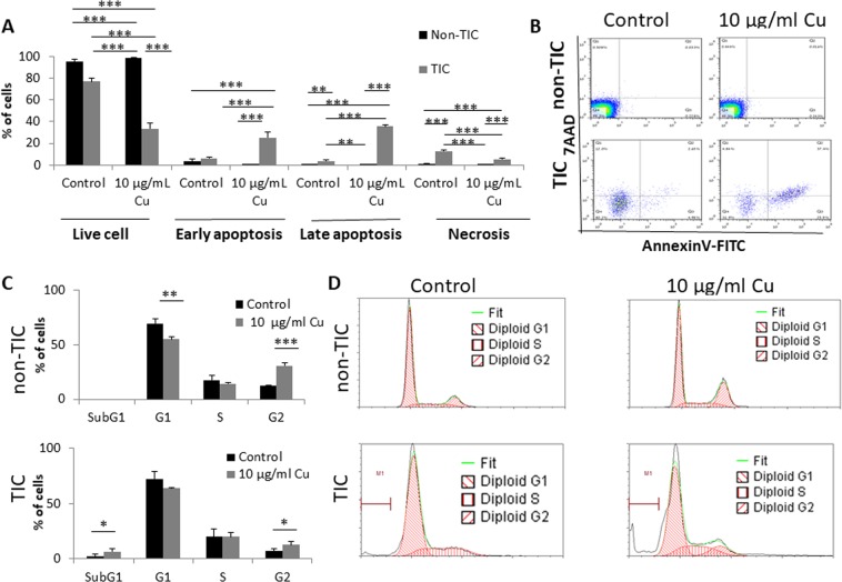
Figure 2.
CuO-NPs induce apoptosis of TICs and promote their arrest in Sub-G1 phase. (A,B) PANC1 cells were grown under standard culture conditions (non-TIC) or TIC-enriching conditions (TIC) in the presence of CuO-NPs (10 µg/ml Cu) for 24 hours. Different stages of cell apoptosis and necrosis were evaluated by 7AAD and Annexin V staining followed by flow cytometry acquisition and analysis. Percentages of cells in each stage are shown in (A), and representative flow cytometry plots are shown in (B). (C,D) PANC1 cells were treated as in (A,B). Cell cycle phases were assessed by flow cytometry. Percentages of cells in each phase are shown in (C), and representative flow cytometry histograms are shown in (D). Experiments were performed in triplicate, with at least two biological repeats. *p < 0.05; **p < 0.01; ***p < 0.001 as assessed by one way ANOVA followed by Tukey post-hoc test.

