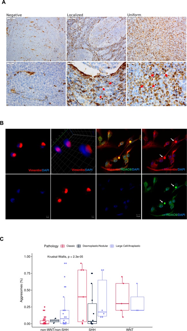
Figure 1.
Characterization of aggresomes in pediatric medulloblastoma tumor samples. (A) IHC analysis of vimentin identified three different patterns. Negative; vimentin immunoreactivity was absent or only observed in infiltrating blood vessels. Localized; observed in desmoplastic/nodular variant, where juxta-nuclear dot-like vimentin was identified in the interstitial space between nodules. Uniform distribution; of juxta-nuclear dot-like were identified in classic and LC/A histological subtypes. Red arrows show para nuclear localization of vimentin indicative of aggresomes formation. (B) IF analysis of CCHE-188 cells. Cells were immunostained with mouse anti-vimentin and rabbit anti-HDAC6 then visualized using Alexa Fluor 488 goat anti-mouse and Alexa Fluor 555 goat anti-rabbit respectively. Cells were counter-stained using DAPI. (C) Bar Plot of the distribution of aggresomes percentages among different molecular subgroups, subdivided according to histological variant. p-values were calculated using Kruskal-Wallis test.

