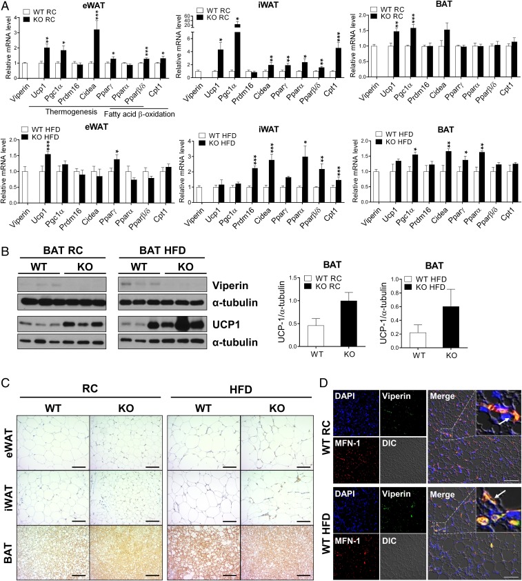
Fig. 2.
Viperin deficiency promotes expression of thermogenesis-related genes and proteins in adipose tissues. The adipose tissues are isolated from WT and viperin KO mice fed RC or an HFD for 15 wk. (A) Relative mRNA levels of thermogenesis- and fatty acid β-oxidation–related genes in adipose tissues (n = 6). (B) Protein expression of viperin and UCP1 in BAT (n = 3). (C) Immunohistochemical staining for UCP1 in adipose tissues. (Scale bar: 200 μm.) (D) Immunofluorescence staining of adipose tissues. DAPI, nucleus (blue); viperin (green); MFN-1, a mitochondrial marker (red). Arrows indicate viperin localized to the mitochondria. (Scale bar: 50 μm.) Data are presented as mean ± SEM of biologically independent samples. *P < 0.05; **P < 0.01; ***P < 0.001 vs. WT on the same diet.

