Fig. 1.
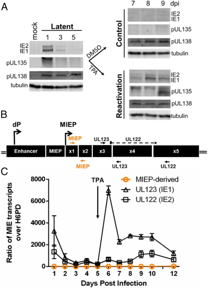
IE1 and IE2 are expressed during HCMV reactivation but do not arise from the MIEP. (A) THP-1 cells were infected with TB40/E WT HCMV (MOI = 2) and cultured for 5 d to allow the establishment of latency. At day 5, cells were treated with TPA or a DMSO control. Whole-cell lysates were collected at the indicated time points, and IE1, IE2, pUL135, and pUL138 viral proteins were detected by immunoblotting. Tubulin was used as a loading control. A single experiment (representative of 3 independent experiments) is shown. (B) Schematic of the major immediate early (MIE) locus. The distal promoter (dP) and major immediate early promoter (MIEP) and primers to detect UL123 (IE1) (exons 3 and 4), UL122 (IE2) (spanning exons 3 to 5), or MIEP/dP-derived transcripts (exons 1 and 2, orange) are indicated. (C) UL123 (IE1), UL122 (IE2), and MIEP/dP-derived transcripts were detected by RT-qPCR over a time course following infection and reactivation in THP-1 cells. Transcripts are quantified as a ratio over H6PD. Data from 3 independent biological replicates (each performed in triplicate) are shown; error bars indicate SE.

