Fig. 3.
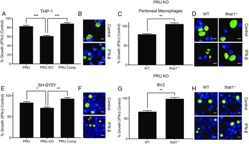
Type I IFN-mediated growth suppression of TgIST-deficient parasites. Comparison of vacuolar growth in control or IFN-β–treated cells of various types infected with type II wild-type parasites (PRU), ∆Tgist knockout (PRU KO), or complemented lines (PRU Comp). Differentiated THP-1 human macrophages (A and B) and SH-SY5Y human neuroblastoma cells (E and F) are shown. ∆Tgist knockout (PRU KO)-infected (C and D), wild-type (WT), Ifnar1−/− thioglycolate-elicited peritoneal macrophages (G and H), mouse microglial WT, and Stat1−/− BV2 cells are shown. Cells were infected for 2 h and then treated with TNF-α (10 ng/mL; Control) alone or in combination with IFN-β (100 units/mL) during 40 h of infection. Growth or mean vacuolar size per image of parasites upon IFN-β treatment relative to control is plotted as % Growth ± SEM of at least 3 independent replicates with at least 50 images per replicate and sample. There were significant differences between the compared samples (*P < 0.05, **P < 0.01, and ***P < 0.001 using 1-way ANOVA in A and E and an unpaired Student’s t test in C and G). Representative images of vacuolar growth of PRU KO parasites in THP-1, peritoneal macrophages, SH-SY5Y, and BV2 cells are shown in B, D, F, and H, respectively. Parasites were stained with mouse mAb anti-SAG1 (green), and the vacuolar membrane was stained with Pc rabbit anti-GRA7 (red) and detected with appropriately conjugated secondary antibodies. Nuclei were stained with Hoechst (100 ng/mL). (Scale bars, 10 μm.)

