Fig. 4.
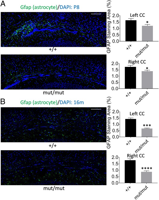
Reduced astrocyte staining in the CC of Gnptab Ser321Gly mouse brains. Perfusion-fixed coronal cryosections (10-µm thickness) were used for immunostaining using anti-Gfap staining for astrocytes in the CC area. Quantification of the stained area was done using ImageJ software and paired t tests were used to test statistical significance of staining differences between genotype groups by calculating 2-tailed P values. (A) Immunostaining and quantitation of the stained area in P8 mice. Sample sizes (n) of +/+ = 10, n of mut/mut = 10, DF is 9 for both hemispheres. (B) Immunostaining and quantitation of the stained area in 16-mo-old mice. The n of +/+ = 8, n of mut/mut = 8, DF is 7 for left hemispheres. The n of +/+ = 9, n of mut/mut = 8, DF is 8 for right hemispheres. (Scale bars, 100 µm.) Error bars indicate the SEM. *P ≤ 0.05; ***P ≤ 0.001; ****P ≤ 0.0001.

