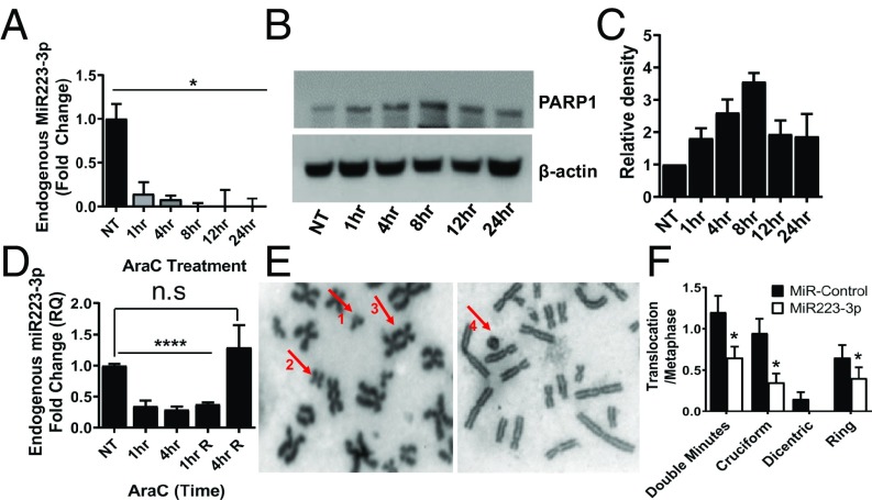
Fig. 2.
MiR22-3p represses chromosomal translocation in hematopoietic cells. (A) QRT-PCR showing the endogenous levels of miR223-3p at different time points after Ara-C treatment in HL-60 cells. (B) Western analysis showing the PARP1 protein levels at different time points after Ara-C treatment in HL60 cells. (C) Quantitation of the Western blots showing relative levels of PARP1 in HL-60 cells after Ara-C treatment. (D) QRT-PCR showing levels of endogenous miR223-3p in HL-60 cells at different time points after release from Ara-C treatment. (E) Representative confocal metaphase images showing chromosomal translocation phenotypes in Jurkat cells after VP16 exposure (1- Double minutes, 2- Cruciform structure, 3- Dicentric chromosomes, and 4- Ring chromosome). (F) Percentage of cells showing different chromosomal translocation phenotypes in Jurkat cells treated with VP16 with or without prior transfection of miR223-3p.

