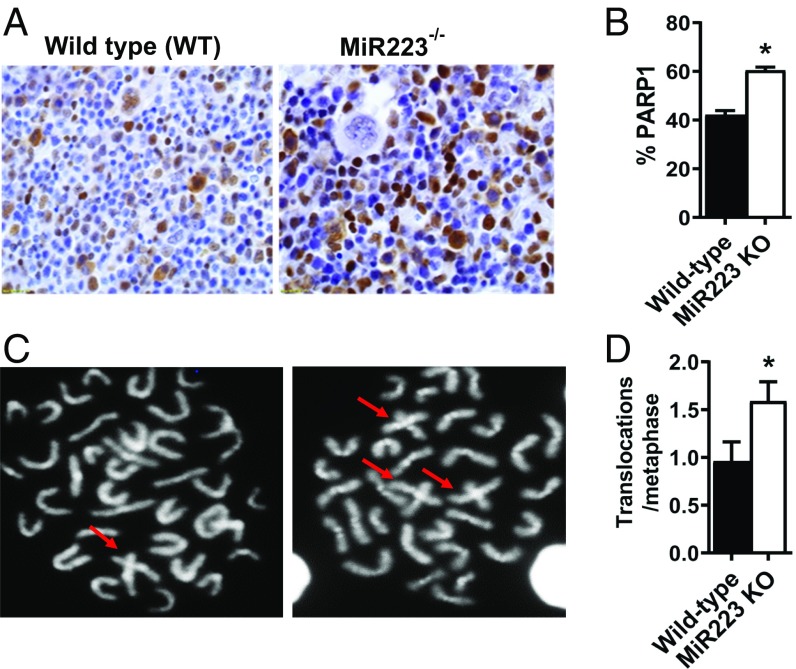
Fig. 3.
MiR223 KO mice exhibit increased unprovoked chromosomal aberrations. (A) Representative images for PARP1 protein assessed by immunohistology in the bone marrow of miR223 wild-type (WT) and genetically deleted mice. (B) Percentage of PARP1-expressing cells in the bone marrow of miR223−/− and WT mice. (C) Representative confocal images showing metaphase chromosomes in the MiR223−/− and WT mouse bone marrow. Cruciform structures indicative of chromosomal fusions are shown with arrows. (D) Percentage of chromosomal aberrations per metaphase in MiR223−/− and WT mice.

