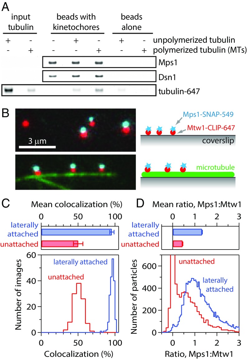
Fig. 1.
Isolated kinetochores retain Mps1 when attached laterally to microtubules. (A) Kinetochores isolated (from SBY9190) and bound to magnetic beads were mock-treated, incubated with unpolymerized fluorescent tubulin, or incubated with fluorescent Taxol-stabilized microtubules (MTs). The amount of tubulin retained after bead washing was assessed by fluorescence imaging. Kinetochores (represented by Dsn1) and kinetochore-associated Mps1 were detected by immunoblotting. (B) Fluorescence images of individual kinetochore particles (from SBY15285) carrying Mps1-SNAP 549 (cyan) and Mtw1-CLIP 647 (red), tethered to a coverslip (Top) or attached laterally to a microtubule (green; Bottom). Colors are offset vertically; cyan-red pairs are colocalized, dual-color particles. (C) Percentages of kinetochore particles retaining Mps1. Bars show mean ± SD values from 192 images of microtubule-attached particles and 112 images of coverslip-tethered particles. Histograms show corresponding distributions. (D) Approximate ratios of Mps1 to Mtw1 molecules, estimated from particle brightness relative to the brightness of single Mps1-SNAP 549 and Mtw1-CLIP 647 molecules. Bars show mean ± SEM ratios from 14,189 laterally attached particles and 7,830 coverslip-tethered particles. Histograms show corresponding distributions.

