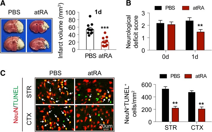
Fig. 1.
Prophylactic atRA treatment protected against acute ischemic stroke. Male C57BL/6 mice were treated with atRA (1 mg/kg, i.p.) or PBS 24 h before 60 min of cerebral ischemia. The treatment was repeated immediately after reperfusion. Mice were sacrificed at 1 day after tMCAO. a Infarct volume of mice was quantified with TTC (red)-stained coronal sections (N = 11 mice for PBS-treated group and N = 10 mice for atRA-treated group). ***P ≤ 0.001, versus PBS-treated group in t test. b Neurological deficit score was assessed right after reperfusion and 1d after tMCAO (N = 11 mice for PBS-treated group and N = 10 mice for atRA-treated group). **P ≤ 0.01, versus PBS-treated group in two-way ANOVA. c Left: Representative images showing TUNEL (green) co-labeling with NeuN (red) in infarct penumbra at 1d after tMCAO. Right: Quantification of the number of NeuN+TUNEL+ neurons (yellow, emphasized with white arrows) in stroke penumbra of striatum (STR) and cortex (CTX). N = 6 mice per group. **P ≤ 0.01, versus PBS-treated group in t test

