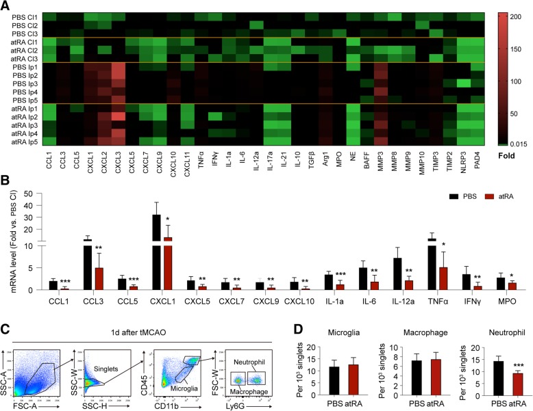
Fig. 2.
Prophylactic atRA treatment attenuated post-stroke neural inflammation. Post-stroke neural inflammation at acute phase after cerebral ischemia was analyzed with qPCR (a, b) and flow cytometry (c, d) at 1 day after tMCAO. a Heat map of the mRNA expression of inflammatory mediators in contralateral brain (Cl) and ipsilateral hemisphere (Ip). In the heat map, data are displayed as fold change to Cl of PBS-treated group. Black, expression un-altered, fold change = 1; Red, expression up-regulated, fold change > 1; Green, expression down-regulated, fold change < 1. b Quantification of inflammatory markers with significant difference of mRNA expression in the Ip brains between atRA- and PBS-treated mice. Comparison between PBS Cl and atRA Cl groups was displayed in Additional file 1: Figure S1A. Quantification of markers that without significant difference between PBS Ip and atRA Ip groups was displayed in Additional file 1: Figure S1B. mRNA expression was normalized to the level of the Cl brain from PBS-treated mice. N = 3 in PBS Cl and atRA Cl groups, N = 5 in PBS Ip and atRA Ip groups. *P ≤ 0.05, **P ≤ 0.01, ***P ≤ 0.001, versus PBS Ip in t test. c Gating strategy for microglia (CD11b+CD45int), macrophage (CD11b+CD45hiLy6G−), and neutrophil (CD11b+CD45hiLy6G+) in flow cytometric analysis. d Quantification of microglia, macrophages and neutrophils among single brain cells (singlets). The number in flow panels represents the number of cells per 103 singlets in ipsilateral brain. N = 6 mice per group. ***P ≤ 0.001 versus PBS-treated group in t test

