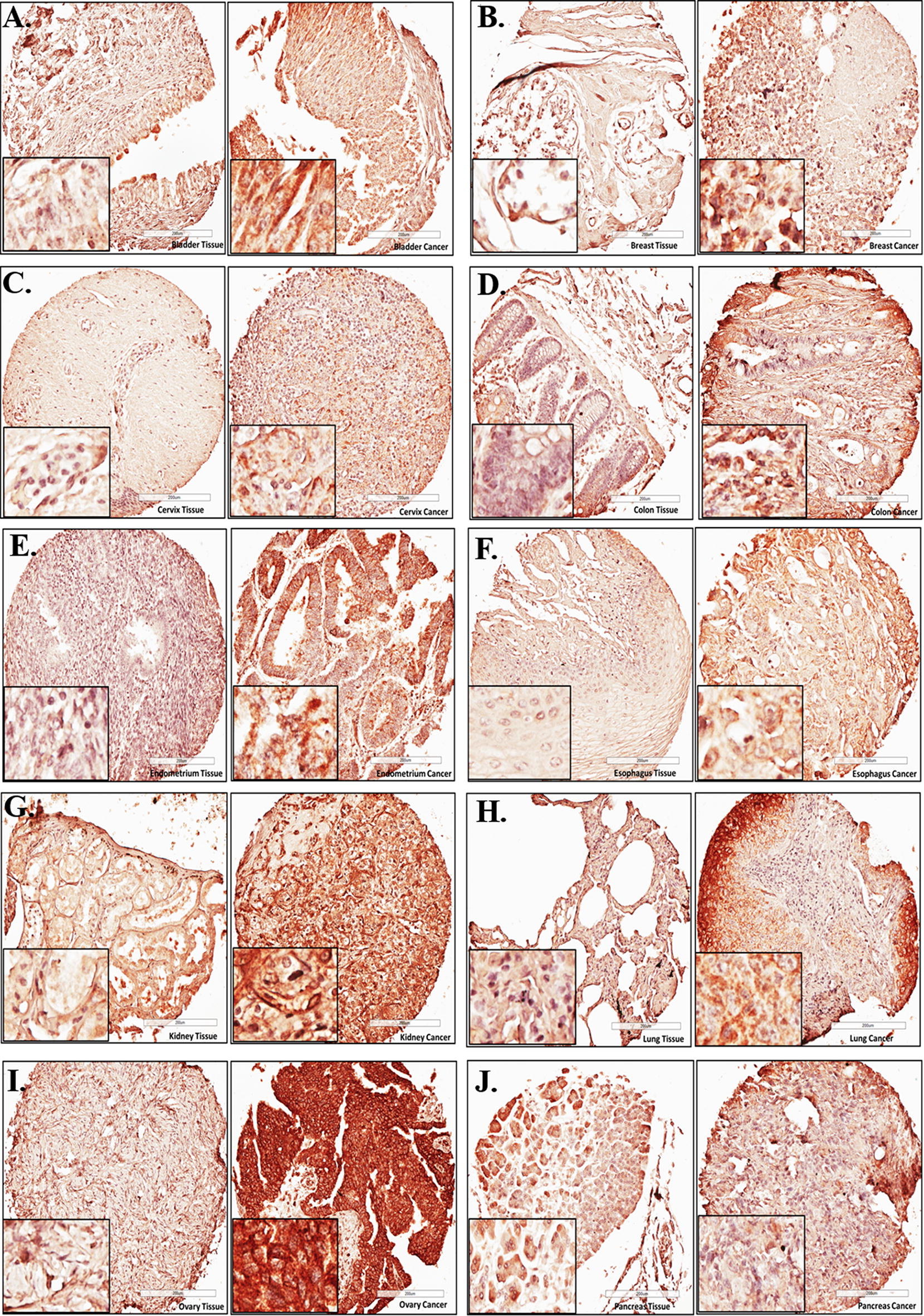
Fig. 1.

High protein levels of EIF4G1 were observed in the tissue sections from different cancers: Representative photomicrographs for EIF4G1 IHC for A bladder, B breast, C cervical, D colon, E endometrial, F esophagus, G kidney, H lung, I ovarian, j pancreatic cancer patients (right side) with respective normal tissues (left side). High-density TMA was scanned through Leica Biosystems CS2 scanner. Aperio ImageScope viewer was used to take photomicrographs for representative tissue sections of IHC
