Fig. 3.
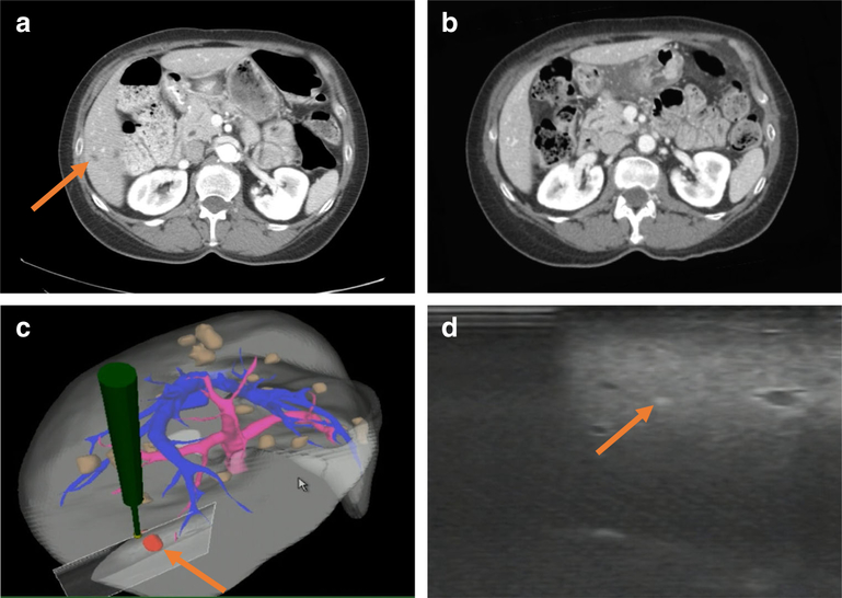
Image guidance system in use. a Last preoperative CT scan where DLM (orange arrow) was radiographically visible, 6 months before date of surgery. b Corresponding section of immediately-preoperative CT scan where DLM has become radiographically occult. c Intraoperative mapping of area of intraoperative US exam to 3D model of the liver with DLM (orange arrow). d Successful identification of DLM (orange arrow) on intraoperative US with image guidance

