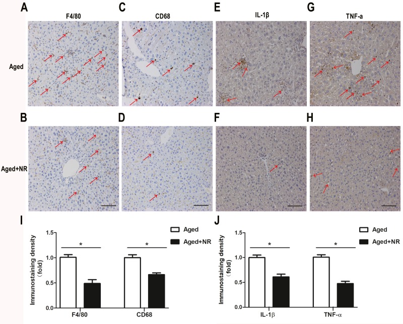
Figure 4. Effect of NR repletion on inflammatory infiltration of aging-relative NAFLD model.
(A–D) Immunohistochemistry staining in liver of aged mice showed the effect of NR supplementation on Kupffer cell accumulation and (I) quantittive analysis. Red arrows show positive brown staining for F4/80 or CD68. (E–H) Protein levels of IL-1β and TNF-α were detected by immunohistochemistry and (J) quantittive analysis. Red arrows show positive brown staining for IL-1β or TNF-α. 200×; Scale bars, 100 µm. Values are mean ± SEM, (n = 5–6 per group). ∗P < 0.05.

