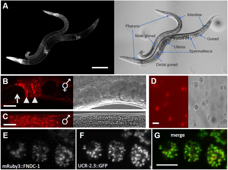
Figure 1 – FNDC-1 is expressed in spermatids and targeted to mitochondria.
mRuby3 red fluorescent protein sequence was inserted into the N-terminal genomic coding region of fndc-1 using CRISPR-Cas9 editing and HDR.
Images in panels A - D contain both fluorescence (left) and differential contrast interference (right).
A Intestine, body wall muscles, and pharynx of males and hermaphrodites, as labeled. Scale bar: 100 μm.
B Hermaphrodite spermatheca. Note the lack of expression in oocytes (arrows) compared to spermatids (arrowheads). Scale bar: 20 μm.
C Male gonad. Scale bar: 20 μm.
D Isolated spermatids from males expressing mRuby3::FNDC-1. Scale bar: 5 μm.
E Higher magnification image of mRuby3::FNDC-1 in spermatids.
F Genomic single copy CRISPR-Cas9 modified ucr-2.3::GFP in the same spermatids as panel E.
G Overlay of mRuby3::FNDC-1 (red) and ucr-2.3::GFP (green). Scale bar: 5 μm.

