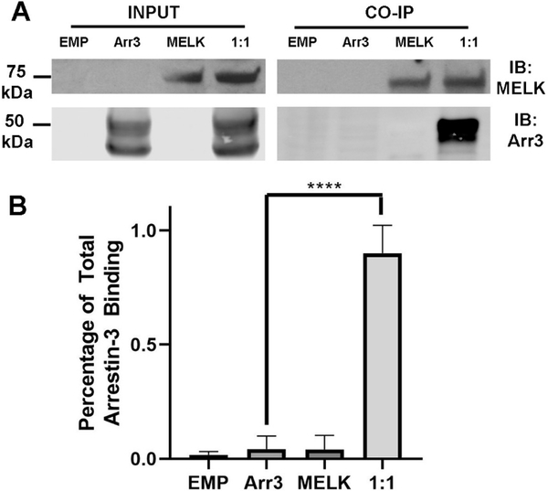
Figure 5. Co-immunoprecipitation of wild-type MELK1−651 with arrestin-31–393.
(A) Western analysis of coimmunoprecipitation. HEK293 arrestin-2/3 knockout cells were transfected with empty vector (20 μg), HA-arrestin-31−393 (10 μg), FLAG-MELK1−651 (10 μg), or both plasmids encoding arrestin-3 and MELK at a 1:1 DNA ratio for 48 h prior to immunoprecipitation with FLAG primary antibody. Western analysis was performed using anti-arrestin (1:10,000; F431) and anti-FLAG (1: 1,000; F3165 Sigma) antibodies. (B) Arrestin-31−393 co-immunoprecipitation with MELK1−651 was quantified using densitometric analysis of arrestin-3 blots. Empty vector and non-specific arrestin-3 binding (no bait control) are also shown. Statistical analysis was performed using One-way ANOVA followed by Dunnett’s post hoc test with correction for multiple comparisons (****, p<0.0001).

