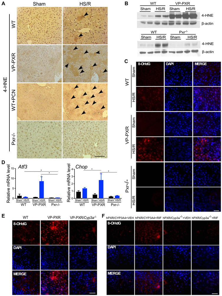
Figure 5. The sensitizing effect of PXR on HS-induced hepatic injury is accompanied by increased oxidative stress.
(A) Immunostaining of 4-HNE in liver tissues from WT, VP-PXR, WT+PCN, and Pxr−/− mice subject to the sham surgery or HS/R. Arrowheads indicate positive staining. (B) The level of 4-HNE was measured by Western blotting. (C) Immunofluorescent staining of 8-OHdG in liver tissue from WT, VP-PXR and Pxr−/− mice. (D) The hepatic expression of Atf3 and Chop was measured by qRT-PCR. (E and F) Immunofluorescent staining for 8-OHdG in liver tissues from WT, VP-PXR and VP-PXR/Cyp3a−/− mice that were subject to HS/R (E), or VEH- or RIF-treated hPXR/CYP3A4 and hPXR/Cyp3a−/− mice that were subject to HS/R (F). Bars are 100 μm. n=4~5 for each group. *, p < 0.05; **, p < 0.01, the comparisons are labeled.

