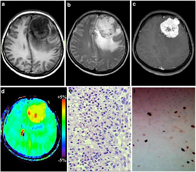
Fig. 4:
A 49-year-old female with atypical meningioma (WHO grade II). a-c The mass was located in the left frontal, it exhibiting hypointense on T1WI, hyperintense on T2WI and obvious heterogeneous enhancement, d On APTw image, the mass exhibiting relative homogeneous signal and the tumor identified approximately equal as on the Gd-Ti weighted images the APTwmax= 4.02%, APTwmin= 3.11% APTwmax-min= 0.71%, APTwmean= 3.87%). Necrosis-related image artefact (Black arrow) and ventricle-related image artefact (White arrow) can be seen.e HE staining showed high nuclear to cytoplasmic ratio cells are arranged loss of whirling or fascicular architecture, f Ki-67 labeling index was 10%).

