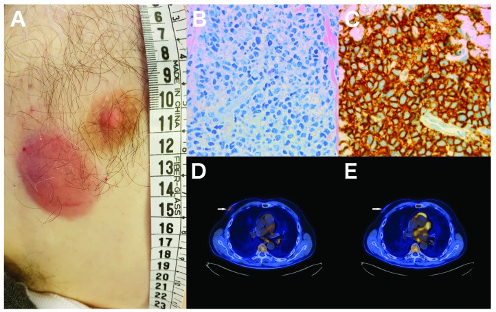
Figure 1.
Clinical morphology, histochemistry, immunohistochemistry and PET-CT of the skin lesion. (A) Clinical photo. (B) Tissue section with medium-sized neoplastic cells with scant cytoplasm. (C) The neoplastic cells show immunoreactivity for CD56. (D) Fused 18F-fluor-deoxy-glucose positron emission tomography/computed tomography (FDG PET/CT) images of the focal skin lesion before the initial treatment with single-agent daratumumab. The focal skin lesion (white arrow) had moderately increased FDG before treatment with a maximum standard uptake value (SUVmax) of 3.2. (E) After the initial treatment the FDG uptake had normalised with a SUVmax of 1.5 and the lesion had reduced in thickness. The patient also had a FDG positive lymph node of the neck (not displayed here) which also reduced in SUVmax from 18.9 to 14.5 and in size from 2.2 × 1.4 cm to 1.7 × 1.0 cm.

