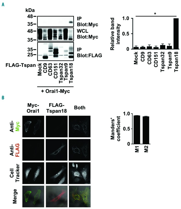
Figure 4.
Tspan18 interacts with Orai1. (A) HEK-293T cells were transfected with a Myctagged human Orai1 expression construct and one of a panel of FLAG-tagged human tetraspanin constructs. Cells were lyzed in 1% digitonin and immunoprecipitated with an anti-FLAG antibody. Samples were separated by SDS-PAGE and both immunoprecipitated (IP) and whole cell lysate (WCL) samples were blotted with anti-FLAG and anti-Myc antibodies. A representative blot for each is shown (left) with quantitation of Myc-tagged Orai1 immunoprecipitated with the tetraspanins (right). Data were nomalized by logarithmic transformation before analysis by one-way ANOVA and Dunnett’s post test. Error bars represent Standard Error of Mean (SEM) from three independent experiments. *P<0.05. (B) HeLa cells were transfected with Myc-tagged human Orai1, FLAG-tagged human Tspan18, or both constructs. Cells were fixed and stained with an anti-Myc antibody (green), an anti-FLAG antibody (red), and imaged by confocal microscopy (left). The Manders’ coefficients (M1 and M2) were calculated from the confocal stacks to quantify the degree of overlap (right). Error bars represent the SEM from three independent experiments.

