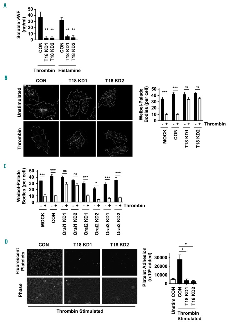
Figure 7.
Tspan18-knockdown endothelial cells have impaired histamine- and thrombin-induced release of von Willebrand Factor (vWF). (A) Human umbilical vein endothelial cells (HUVEC) were transfected with a negative control siRNA (CON) or with one of two independent siRNA targeting Tspan18 (T18 KD). After 48 hours, HUVEC were stimulated with 1 U/mL thrombin or 20 μM histamine for 5 minutes (min). Cultured media was removed and assayed for vWF by ELISA. Pre-stimulation levels of vWF were subtracted from these data. Error bars represent the Standard Error of Mean (SEM) from three experiments. **P<0.01. (B) HUVEC transfected as described in (A) were stimulated with 1 U/mL thrombin for 5 min, and the cells were fixed and stained with an anti-vWF antibody followed by Alexa488-conjugated secondary antibody. Representative confocal microscopy images are shown (left). Z-stack images were de-noised, background-subtracted and analyzed for the number of vWF cellular bodies, using ImageJ. Counts were made on 5-10 cells per experiment for four independent experiments (right panel). Error bars represent Standard Error of Mean (SEM). ***P<0.001; ns: not significant. (C) The experiment and quantitation was conducted as for (B), except that HUVEC were mock-trans-fected with no siRNA (Mock), transfected with negative control siRNA (CON), or siRNA to Orai1, Orai2 or Orai3 (KD). (D) HUVEC were siRNA-transfected and stimulated with 1 U/mL thrombin for 5 min as described in (A). Human washed platelets were fluorescently labeled and incubated with the HUVEC monolayers. Non-adherent platelets were removed by washing and images were collected. Representative fluorescent images of adhered platelets (top) and phase contrast images of the HUVEC monolayers and adhered platelets (bottom) are shown, with quantitation of platelet adhesion from three independent experiments (right). Error bars represent SEM. *P<0.05.

