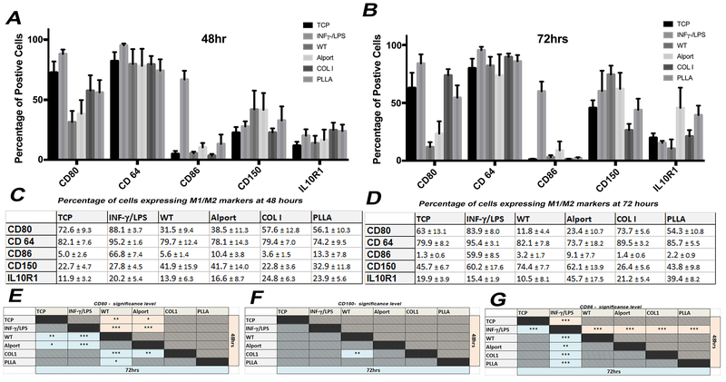
Figure 5: Phenotypical analysis of macrophages seeded on mrECM (WT and Alport) or synthetic biomaterial.
A-B. Graphs representing the percentage of cells positive for CD80, CD64, CD86, CD150 and IL10R1 cultured and seeded in different conditions (non stimulated-TCP, INF-γ/LPS stimulated, WT mrECM, Alport mrECM, collagen I and PLLA) after 48hrs (A) and 72hrs (B). C-D. Table reporting n percentage of macrophages expressing M1/M2 markers for all samples analyzed at 48hrs (A) and 72hrs (B). All values are presented as mean ± SEM. E-G. Tables summarizing statistical significant differences for results represented in A-D between the different groups at 48hrs (light red cells) and 72 hrs (light blue cells) for CD80, CD150 and CD86. No significance was evident for CD64 and IL-10R1 between the experimental groups , *p-value <0.05; **p-value <0.01; *** p-value <0.001.

