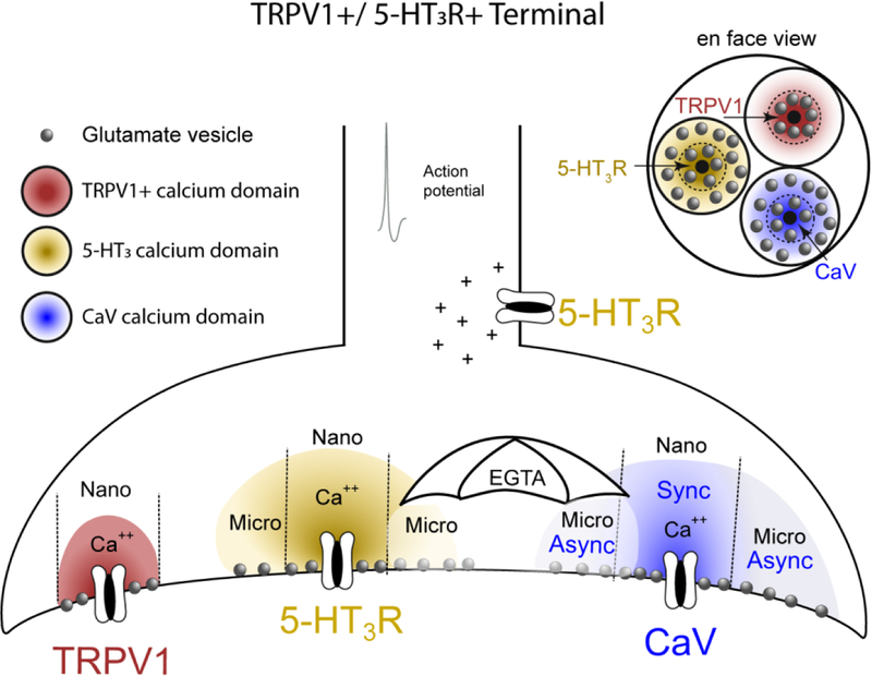
Figure 11.
Schematic illustration of the hypothesized calcium domains and vesicle arrangements of 5-HT3R, CaV and TRPV1 on a single ST afferent terminals. Calcium (Ca++, colored domes) enters the channel and mobilizes glutamate vesicles (gray spheres) at varying distances from the calcium source. Calcium spreads from the channel pore opening exposing the surrounding vesicles to calcium concentrations that decline with distance (represented by the color density gradient). 5-HT3Rs located on the axon slows action potential conduction, often preventing action potentials from invading the synapse. Calcium entry through terminal 5-HT3Rs mobilizes vesicles within both a nanodomain (nano) and microdomain (micro) that does not overlap the calcium domains and respective vesicle pools from other calcium channels. EGTAAM (EGTA, umbrella) defines calcium domains based on its time-dependent ability to chelate calcium. EGTA is ineffective at chelating calcium within an immediate nanodomain close to the pore. EGTA effectively chelates calcium and reduces calcium levels distal to the pore, and reduces release by “shielding” vesicles from release within the microdomain. Each calcium source has a vesicle population located within nanodomains whose release is unaffected by EGTA. 5-HT3R and CaV both have vesicles additionally located within a microdomain but TRPV1 does not.

