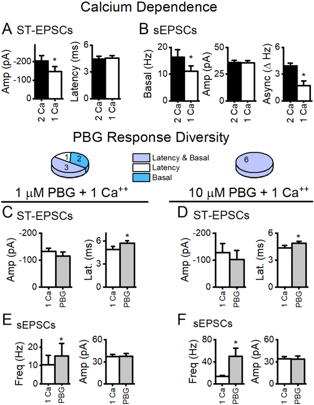
Figure 9.
Reduced external calcium decreased glutamate release but did not change PBG responses. A, The amplitude of ST-EPSCs is calcium-dependent (p < 0.01, paired t test, n = 14) while the latency from the ST shock to the onset of glutamate release remained unchanged (p = 0.2, paired t test). B, Spontaneous glutamate release was also calcium sensitive (p < 0.01, paired t test) and the asynchronous component was strongly reduced in 1 mM Ca ++ (p = 0.01, paired t test). Similar to results in 2 mM external calcium, 1 μM PBG (C, E) and 10 μM (D, F) increased latencies (p = 0.03, n = 5; p = 0.048, n = 4, respectively, paired t tests) but not amplitudes (p = 0.1, n = 3; p = 0.1, n = 5, respectively, paired t tests) of ST-EPSCs and increased frequencies (p = 0.03, n = 6; p < 0.001, n = 5, paired t tests) but not amplitudes of basal sEPSC activity (both p values > 0.8, paired t test). These results demonstrate that reducing the calcium driving force does not change the diversity of PBG responses.

