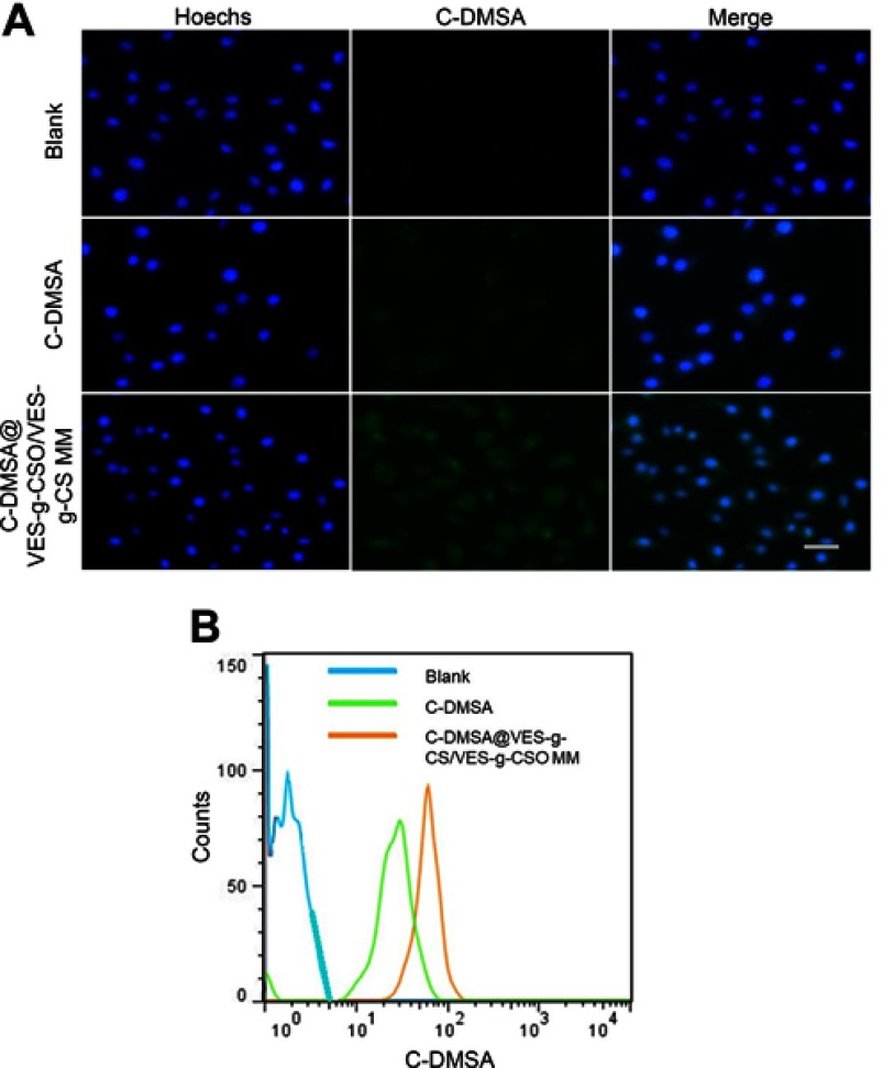
Figure 4.
Fluorescence microscopy images (A) and flow cytometry (B) of L929 cells after incubation with PBS.
Notes: Blue, cell nuclei stained with Hoechst 33258; green, fluorescence of coumarin-aldehyde generated by C-DMSA after its reaction with Hg2+.
Abbreviations: C-DMSA, 3-formyl-7-diethylamino coumarin masked meso-dimercaptosuccinic acid; C-DMSA@VES-g-CSO/VES-g-CS MM, C-DMSA loaded vitamin E succinate-grafted-chitosan oligosaccharide/vitamin E succinate-grafted-chitosan mixed micelles.

